
A
weekly e-journal by Art Epstein, OD, FAAO
|
|
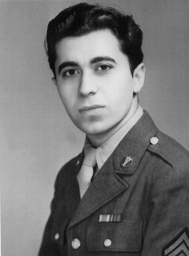
Off the Cuff: A Time of Great Sorrow – The Passing of Uncle Frank Fontana
It is with deep sadness that I note the passing of my dear friend, mentor and role model Uncle Frank Fontana. A St. Louis-based, internationally recognized optometric icon, Frank was an unstoppable force of nature who helped shape the profession, but will be best remembered by his many friends for his incredible humanity. Frank had many “nieces” and “nephews,” and I was proud to be among them. He was the only person I’ve ever met who carried photos of his friends’ kids in his wallet. The title “Uncle” wasn’t honorary. Frank earned it.
|
||||||


|
|||

| Relevance of Swept-Source Anterior Segment Optical Coherence Tomography for Corneal Imaging in Patients With Flap-Related Complications After LASIK | ||||
These researchers evaluated the role of swept-source anterior segment optical coherence tomography (AS-OCT) in the diagnosis and management of laser in situ keratomileusis (LASIK) flap-related complications. This prospective study included 25 eyes with LASIK flap-related complications imaged using swept-source AS-OCT between February and August 2016, at Alforsan Eye Centre, Assiut, Egypt. The images were acquired using a 6-mm line scan. Imaging of flap-related LASIK complications using AS-OCT revealed specific and nonspecific findings. Of note, epithelial ingrowth appeared as highly reflective lesions below the LASIK flap in the form of islands, nests, or a continuous sheet with or without changes in the overlying flap. Macrostriae manifested as dome-shaped irregularities on the stromal surface with regular overlying epithelium, whereas microstriae appeared as corrugations on the stromal surface with regular overlying epithelium. Less common complications included multiple flap macrostriae accompanied by a traumatic folded flap with a flap edge at the interface. Interface debris appeared as a highly reflective interface lesion with or without a surrounding reaction. One eye with a flap that was torn and lost intraoperatively showed epithelialization over a thin residual stroma underlying a contact lens with no stromal infiltration on the second postoperative day. AS-OCT was useful for the assessment of flap thickness and planning of the new flap thickness in the event of an incomplete cut. Researchers wrote that swept-source AS-OCT was useful not only for diagnosis, but also for management of eyes with LASIK flap-related complications, by enabling noninvasive, noncontact, real-time acquisition of cross-sectional AS images. |
||||
SOURCE: Abdelazeem K, Sharaf M, Saleh MGA, et al. Relevance of swept-source anterior segment optical coherence tomography for corneal imaging in patients with flap-related complications after LASIK. Cornea. 2018; Sep 27. [Epub ahead of print]. |
||||
 |
||
| Analysis of Corneal Higher Order Aberrations in Cataract Patients With High Myopia | ||||
Researchers evaluated the differences in corneal higher order aberrations (HOAs) between cataract patients with high axial myopia and normal cataract patients, and identified the associated factors. Corneal aberrations and axial lengths (ALs) were measured using a rotating Scheimpflug camera (Pentacam) and partial coherence interferometry (IOLMaster) in the high myopia group and the control group.
The study included 287 patients (520 eyes): 194 eyes in the high myopia group and 326 eyes in the control group. The five anterior corneal aberrations—vertical coma, vertical trefoil, horizontal coma, oblique trefoil and primary spherical aberration—in the high myopia group were 0.07μm ± 0.38 (SD), -0.11μm ± 0.23μm, 0.07μm ± 0.28μm, -0.02μm ± 0.18μm, and 0.39μm ± 0.19μm, respectively. No negative primary spherical aberrations of the total or anterior corneal surface were found in the high myopia group. Differences between the two groups were found in terms of central corneal thickness, astigmatism, primary spherical aberration, vertical coma and oblique trefoil; however, these differences were not consistent between different age subgroups. Higher order aberrations were correlated with age. Posterior corneal vertical coma was correlated with AL. Negative primary spherical aberrations of the anterior or total corneal surface were not found in the high myopia group. Age showed a strong relationship with HOAs. Investigators wrote that, aspheric intraocular lens implantation is recommended for cataract patients with high myopia. |
||||
SOURCE: Zhang M, Jing Q, Chen J, Jiang Y. Analysis of corneal higher-order aberrations in cataract patients with high myopia. J Cataract Refract Surg. 2018; Sep 28. [Epub ahead of print]. |
||||
 |
||
| Prevalence of Keratoconus in a Refractive Surgery Population | ||||
This study examined the prevalence of keratoconus among patients who were interested in undergoing refractive surgery. Corneal tomography measurements were used to help detect keratoconus. Adult subjects who presented to the private hospital Cataract and Refractive Surgery Unit (Abha, Saudi Arabia) for refractive surgery evaluation were considered for inclusion in this cross-sectional, retrospective study. All subjects were from the Aseer province, a southern, high-altitude region in Saudi Arabia, and presented between January and December 2017. The incidence of keratoconus and other refractive surgery contraindications were examined. A total of 2,931 patients were considered for inclusion in analyses. Of these, 2,280 patients (77.8%) were not candidates for refractive surgery. These 2,280 patients had a mean age of 24.1 years ± 6.6 years and 1,231 patients (54.0%) were male. Of the subjects who did not undergo refractive surgery, 548 (24%) had keratoconus, 400 (17.5%) were keratoconus suspects, 344 (15.1%) had thin corneas, 321 (14.1%) had high myopia and 52 (2.3%) had a high astigmatism. An additional 479 subjects (21%) were candidates for refractive surgery, but chose not to undergo a procedure. The incidence of keratoconus in Saudi Arabian refractive surgery prospects was 18.7%. Keratoconus was the most common reason for not performing refractive surgery and accounted for 24% of cases in which surgery was not performed. |
||||
SOURCE: Al-Amri AM. Prevalence of keratoconus in a refractive surgery population. J Ophthalmol. 2018;2018:5983530. |
||||
|
|||

| News & Notes | |||||||||||||||||||||||||||||
Essilor Launches Pro-E 700 Edging System
|
|||||||||||||||||||||||||||||
| NORA Names Members to Advisory Board The Neuro-Optometric Rehabilitation Association, International announced the addition of six new members to its advisory board at the group’s 27th Annual Conference in St. Louis. Joining the advisory board are: • Tanya Polec, OD, FCOVD, neuro-optometric concussion specialist, VQ Learning Sports Rehab in North Central Tucson, Ariz. • Hannu Laukkanen, OD MEd FAAO FCOVD-A, professor emeritus of optometry, and graduate course instructor, Pacific University’s Vision Science • Jill Schultz, OD, FAAO, in practice, Bright Eyes Vision Clinic in Minnetonka and Otsego, Minn. • Shirley Ha, OD, FCOVD, graduate, University of Waterloo’s School of Optometry and Vision Science; board-certified fellow of the College of Optometrists in Vision Therapy • Patti Andrich, MA, OTR/L, COVT, CINPP, occupational therapist, Vision Development Team, Royalton, Ohio • Gina Kim, MOT, OTR/L, CBIS, lead therapist/occupational therapist, Centre for Neuro Skills, Encino, Calif. Read more. |
|||||||||||||||||||||||||||||
|
|||||||||||||||||||||||||||||


|
Optometric Physician™ (OP) newsletter is owned and published by Dr. Arthur Epstein. It is distributed by the Review Group, a Division of Jobson Medical Information LLC (JMI), 11 Campus Boulevard, Newtown Square, PA 19073. HOW TO ADVERTISE |